- nulifediagnostics2023@gmail.com
- 5303 Yarmouth Ave # 104 Encino, CA 91316
Ultrasound is one of the least invasive and most widely used diagnostic medical tools available. Compared to other procedures, an ultrasound study generally involves no discomfort and requires very little patient preparation.
Ultrasound transmits safe, non-invasive, ultra-high frequency sound waves and creates an image from the resulting echoes. These echoes are recorded, processed, and displayed on a screen by a computer.
Unlike X-ray, ultrasound does not require the use of radiation, and it does not focus on bone structures. Rather, ultrasound is used to study internal organs, such as the heart, liver, uterus, ovaries, blood vessels, and other soft tissue structures.
During your ultrasound exam, our Sonographer will position you on an exam table, apply a topical gel to the skin (this helps to improve the quality of the images), and pass the transducer several times over the area to be examined.
Depending on the type of study being performed, you may be required to remain motionless, change positions and/or hold your breath.

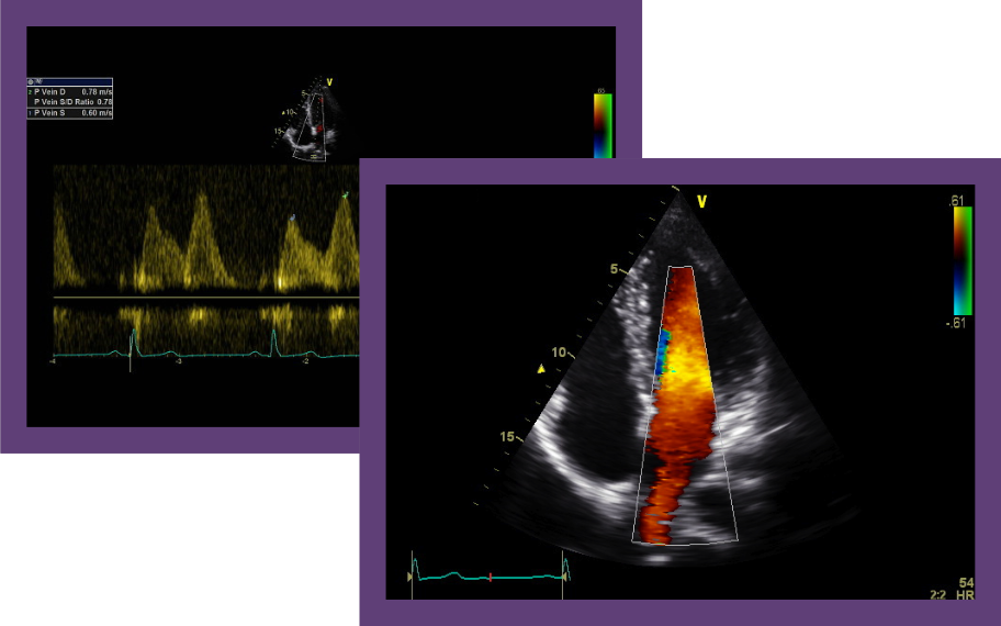
The person who will perform your exam is a medical professional known as a Sonographer. Our Sonographers are some of the most highly trained and experienced professionals in the country. Each Sonographer is certified by the American Registry of Diagnostic Medical Sonographers (ARDMS®).
A full report detailing the findings and interpretation of the results will be provided to your physician. For compliance reasons, our Sonographers cannot discuss the study findings with you until your physician has the final report. Your physician will discuss the ultrasound findings with you after reviewing the final diagnostic report.
Your ultrasound will be performed right in the comfort of your physician’s office. Nulife Diagnostics’ on-site service means convenience for you.
Nulife Diagnostics on-site service offers you the benefit of reduced patient cost. Our diagnostics are cost-effective, saving you from facility fees and specialty co-pays that are associated with outside imaging centers.
| Ultrasound Exam |
Exam Time (in minutes) |
Patient Preparation & Dietary Restrictions |
| Abdominal | 30 | Please do not eat or drink anything 6-8 hours prior to the exam. Avoid fatty foods and carbonated liquids the day prior to your exam. |
| Pelvic | 30 | No dietary restrictions, except you must complete drinking 32 oz. of water one hour prior to the exam. You should not empty your bladder once you have started drinking. |
| Abdominal & Pelvic | 60 | Avoid fatty foods and carbonated liquids the day prior to the exam. You must complete drinking 32 oz. of water one hour prior to the exam. You should not empty your bladder once you have started drinking. |
| Renal Artery Duplex | 30 | Please do not eat or drink anything 6-8 hours prior to the exam. Avoid fatty foods and carbonated liquids the day prior to your exam. |
| Abdominal Aorta/IVC Duplex | 30 | Please do not eat or drink anything 6-8 hours prior to the exam. Avoid fatty foods and carbonated liquids the day prior to your exam. |
| Echocardiogram | 45 | No patient preparation required. |
| Renal/Retroperitoneal | 30 | No dietary restrictions, except you must complete drinking 32 oz. of water one hour prior to the exam. You should not empty your bladder once you have started drinking. |
| Carotid Duplex | 30 | No patient preparation required. |
| Upper/Lower Extremity Venous Duplex | 30 | No patient preparation required. |
| Upper/Lower Extremity Arterial Duplex | 30 | No patient preparation required. |
| Testicular/Scrotal | 30 | No patient preparation required. |
| Thyroid | 30 | No patient preparation required. |
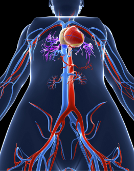
The Upper or Lower Extremity Venous Ultrasound is a diagnostic test used to check the circulation in the large veins in the legs or the arms. This exam shows any blockage in the veins by a blood clot or thrombus with compression of the veins. It can also help to diagnose venous insufficiency in the legs which can cause varicose veins. Venous Ultrasound uses real time imaging and color Doppler to evaluate the venous system. It takes less than 30 minutes and there is no patient preparation required for a venous ultrasound.
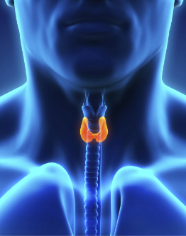
The thyroid is a butterfly-shaped gland that wraps around the front part of the neck just below your Adam’s apple. The thyroid makes hormones that control the body’s metabolism. The hormones produced by the thyroid have an effect on body temperature, digestive and heart functions, and other body processes. Thyroid Ultrasound is an effective tool that takes less than 30 minutes to assist in the diagnosis of thyroid disease.

The scrotum contains the male reproductive organs consisting of the vas deferens, epididymis, pampiniform plexus, spermatic cord, and the testicles. Ultrasound is the gold standard for examining the testicles with real-time imaging. Scrotal Ultrasound is an efficient and effective way to examine the scrotal contents. The exam takes 30 minutes and monitors the venous and arterial blood flow. Gray scale ultrasound imaging identifies testicular disease or malformations.
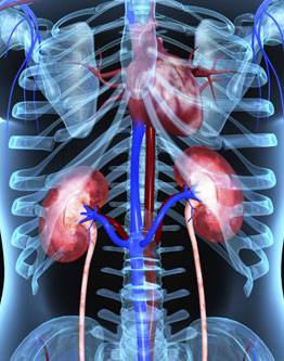
The Renal Artery Duplex Ultrasound study is an accurate, non-invasive and cost-effective way in which to examine the function of the kidneys. The kidneys play an important role in regulating blood pressure by secreting a hormone called renin. If the renal arteries are narrowed or blocked, the kidneys are unable to effectively control blood pressure. Persistent or severe high blood pressure is a common symptom of renal artery stenosis. The Renal Artery Duplex Ultrasound study provides a real-time image of blood flow and accurate velocities of the blood to the renal artery. A Renal Artery Duplex Ultrasound can be performed with no radiation exposure, unlike a CT scan, and at a significantly lower cost than an MRI exam.

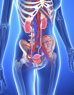
A Pelvic Ultrasound is an efficient and cost-effective way to assess the pelvic organs, which include the uterus, cervix, vagina, fallopian tubes and ovaries. Doppler is also performed in a Pelvic Ultrasound to show blood flow to the major reproductive organs. During a Pelvic Ultrasound, the bladder is also visualized. Pelvic Ultrasound utilizes real-time imaging and color Doppler to rule out pelvic disease or abnormalities. The exam takes 30 minutes to perform and is best done with the patient prepped with a full bladder.

An Echocardiogram is an ultrasound California of the heart that tests its beating, pumping, and blood flow. Echocardiograms are a wonderful tool for you and your patient and can be used in the diagnosis of valvular stenosis or sclerosis, valvular regurgitation, congenital heart disease, pulmonary hypertension, cardiomyopathy and pericardial effusion.
Heart disease is the leading cause of death for men and women in the United States. Ultrasound in Onpoint Sonography Inc is a safe, effective tool that can be used to prevent heart disease for your practice.

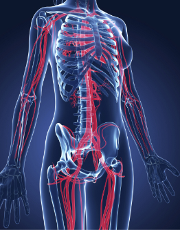

Abdominal Ultrasound provides real-time images of the major organs of the body. Abdominal Ultrasound may be used to diagnose abdominal aortic aneurysms, renal masses, gallstones, pancreatic masses, liver disease, splenic infections or common bile duct obstructions.
An Abdominal Ultrasound Testing takes 30 minutes and must be performed on a fasting patient. Patients should not eat or drink 6-8 hours before the exam.